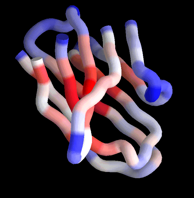
The VH core coloured according to structural variability. Blue are highly variable regions of structure and red are highly conserved. This picture was generated from a multiple-structural alignment of 24 antibody molecules. From the alignment, an average core was computed, and the colors actually show the order in which atoms were "thrown out" in the computation of the core.
For a view of the same core using ellipsoids, click here.
24 January 1994 / mbg@hyper.stanford.edu